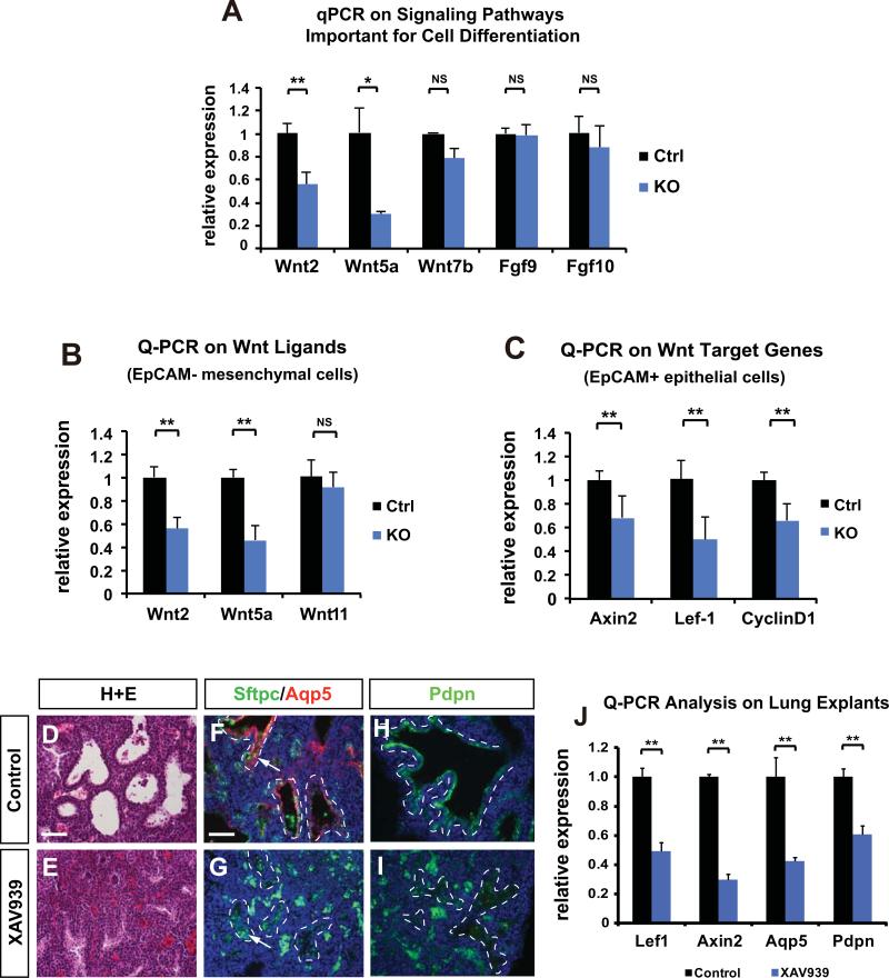
Figure 4. Inhibition of Wnt/β-catenin signaling phenocopies the effect of loss of mesenchymal HDAC3 on AT1 cell differentiation and lung sacculation.
(A) HDAC3Dermo1creKO lungs exhibit decreased expression of several Wnt ligands as assessed by Q-PCR on whole lungs at E18.5. (B) Q-PCR analysis shows that Wnt ligands including Wnt2 and Wnt5a are significantly decreased in the mesenchymal cells of HDAC3Dermo1creKO lungs at E18.5. (C) Target genes of Wnt/β-catenin pathway are decreased in the epithelial cells of HDAC3Dermo1creKO lungs at E18.5. (D-E) Treatment of Wnt/β-catenin signaling inhibitor XAV939 on E16.5 lung explants for 48hrs results in lung sacculation defects as shown by H&E staining. (F-I) Wnt inhibition also leads to a defect in AT1 cell differentiation as shown by the reduction of Aqp5 and Pdpn staining (arrows). Dotted lines outline the boundary between airway epithelium and mesenchyme. Of note, most of the green staining outside of the airways is due to background autofluorescence from red blood cells. (J) XAV939 treatment reduced expression of the canonical Wnt signaling target genes Axin2 and Lef1 and the AT1 cell markers Aqp5 and Pdpn.
Ctrl=Control; KO= Hdac3Dermo1creKO lungs. Two tail student's t test: *p<0.05; **p<0.01; NS=Not Significant. n=6. Q-PCR data are represented as mean ± SD. Scale Bars: 50 μm.

