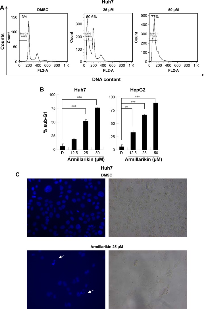
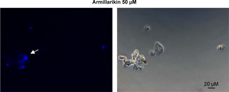
Figure 4.
Armillarikin-induced apoptotic cell death of human HCC cells.
Notes: (A and B) Huh7 and (B, right panel) HepG2 cells were treated with DMSO or armillarikin for 3 days, and the percentage of apoptotic cells (hypodiploid cells) with DNA ladders was detected using flow cytometry. **P<0.01 and ***P<0.001 (Student’s t-test) compared with DMSO control. (C) Armillarikin-induced micronuclei morphology. After Huh7 cells had been treated with armillarikin for 3 days, they were fixed and stained with Hoechst. Fluorescent and cell images were captured; arrow heads indicate micronuclei 400×.
Abbreviations: DMSO, dimethyl sulfoxide; HCC, hepatocellular carcinoma.


