. 2014 Oct 24;25(3):269–281.

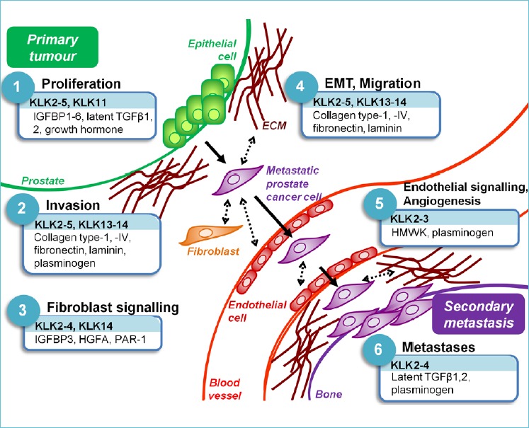
Figure 1.

KLKs implicated in 6 ‘hallmarks’ of prostate cancer progression, by their in vitro substrates, are shown. KLK-expressing prostate cancer epithelial cells exhibit deregulated proliferation and invade through the surrounding basal lamina and extracellular matrix (ECM). Cancer cells interact with stromal fibroblasts mroblasts, as well asundergoing anepithelial-to-mesenchymal transition migratory prostate cancer cells interact with endothelial cells, lining blood vessels and gain entry to the vasculature. Cancer cells must also activate blood vessel formation or angiogenesis, to supply oxygen and nutrients to the expanding primary tumour. Migrating prostate cancer cells extravasate at the compatible secondary site, bone, whereby they degrade the surrounding matrix and form osteoblastic metastases. Arrows show the physical migration of a prostate cancer cell from the primary tumour to a secondary metastatic deposit. Dotted arrows represent KLK-mediated interactions between prostate cancer cells and cells or ECM in the surrounding microenvironment. Only those substrates referenced in the text are shown. IGFBP, insulin-like growth factor binding protein; TGF, transforming growth factor; HGFA, hepatocyte growth factor activator; PAR, protease-activated receptor; HMWK, high molecular weight kininogen.

The role of KLK-related peptidase (KLK)-mediated proteolysis in prostate cancer progression.