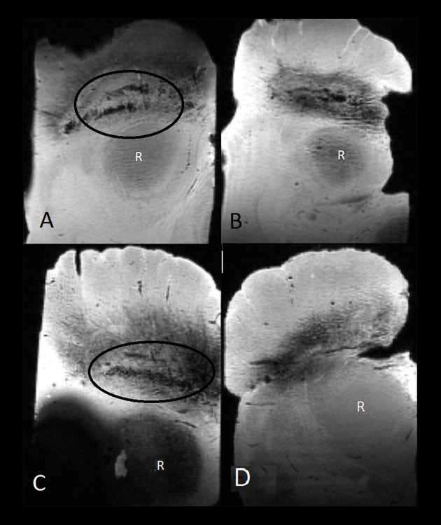
Figure 2.
Ex vivo midbrain samples scanned with a multi-gradient echo sequence on a 11.7-T MRI scanner. A swallow tail appearance of nigrosome-1 is identified in the lower part of the substantia nigra pars compacta (encircled) in two control subjects (A, C). Nigrosome-1 could not be identified in the two samples diagnosed with Parkinson’s disease (B, D). R – red nucleus.

