
Figure 1.
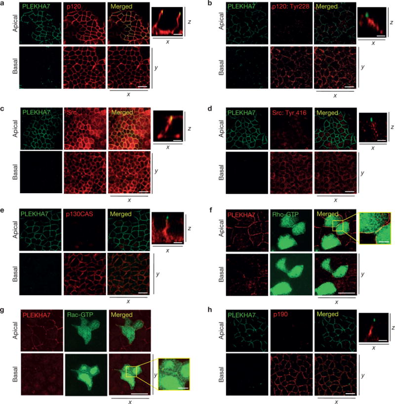
Polarized epithelial cells show distinct p120-associated populations at the junctions. Caco2 cells were grown for 21 days to polarize and subjected to IF for PLEKHA7 and (a) p120, (b) phosphorylated p120 Tyr 228, (c) Src, (d) phosphorylated Src Tyr 416; (e) p130CAS and (h) p190RhoGAP. Also, Caco2 cells were transfected with (f) a green fluorescent protein (GFP)–rGBD (rhotekin RhoA-binding domain) construct to detect active Rho (Rho-GTP) or (g) a yellow fluorescent protein (YFP)– PBD (PAK-binding domain) construct to detect active Rac (Rac-GTP), and co-stained with PLEKHA7. In all cases, stained cells were imaged by confocal microscopy and image stacks were acquired, covering the entire polarized monolayer between the basal and the apical level. Representative x–y image stacks and merged composite x–z images are shown. Enlarged parts of merged images in f and g indicate areas of cell–cell contact. Scale bars for x–y images, 20 μm; for x–z images, 5 μm; for enlarged parts of f and g, 3 μm. PLEKHA7 background staining in f and g is an artefact of paraformaldehyde fixation.