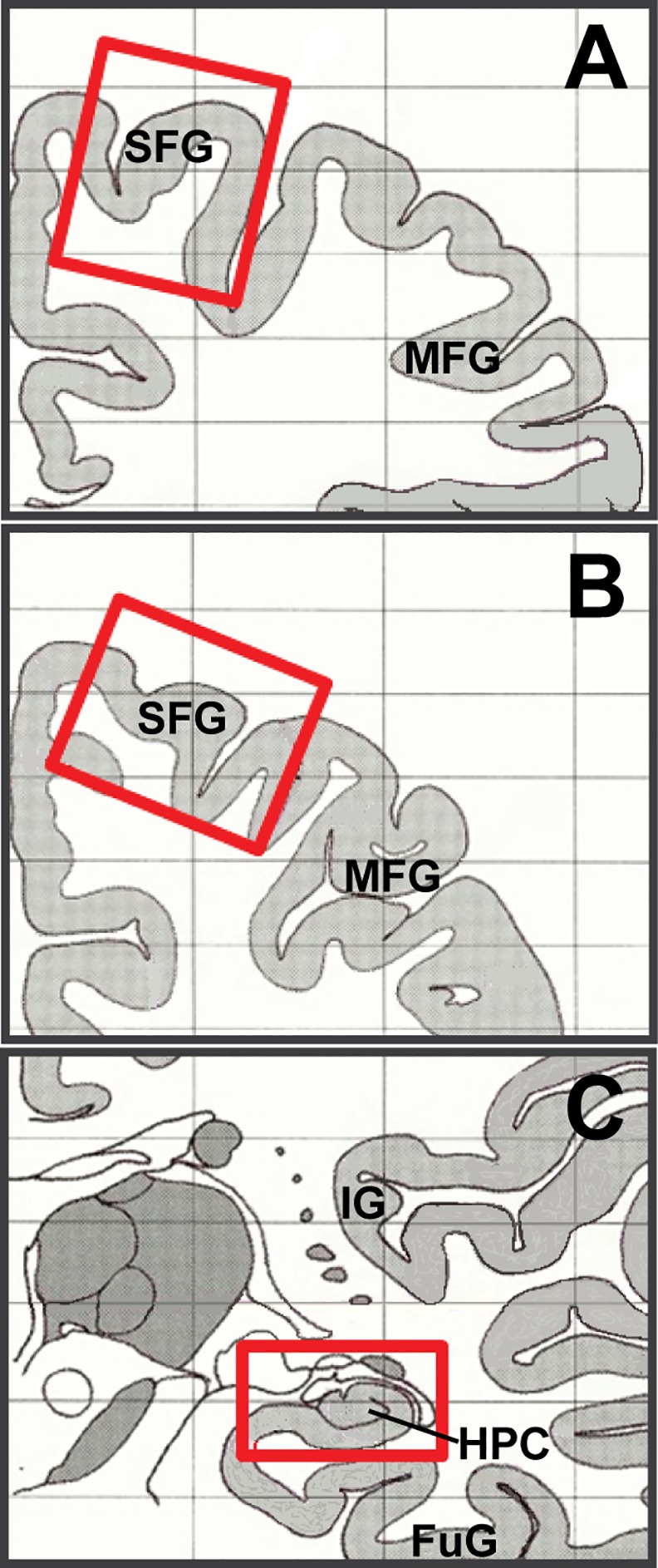
Fig 1. Diagram of dissection regions for human brain samples.

Drawings represent anterior (A) and posterior (B) coronal sections of human brain used for dissection of PFC samples, and (C) HPC samples. Red boxes highlight areas of dissection. SFG: superior frontal gyrus; MFG: middle frontal gyrus; IG: insular gyrus; FuG: fusiform gyrus.
