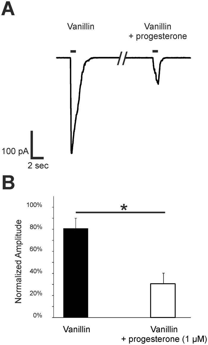
Fig 5. Progesterone decreased vanillin-induced responses in the ORNs of p5 mice.
(A) Representative responses to 100 μM vanillin were obtained from Olfr73 positive ORNs in tissue slices using patch-clamp recordings in a voltage-clamp configuration (Vh = -56 mV). The amplitude was decreased after a 1-min preincubation with 1 μM progesterone. Application time was 1 s as indicated by the application bar. (B) As a control, neurons were challenged with two subsequent applications of 100 μM vanillin. To characterize the effect of progesterone, 100 μM vanillin was applied followed by a progesterone preincubation and a subsequent second application of vanillin. The bar diagram shows the second response with or without progesterone preincubation relative to the first vanillin application. Relative to the second application of vanillin after 1 min in control neurons, the response of Olfr73 positive neurons was significant decreased in neurons preincubated with 1 μM progesterone for 1 min (n = 7). Significant data are labeled: *p ≤ 0.05.

