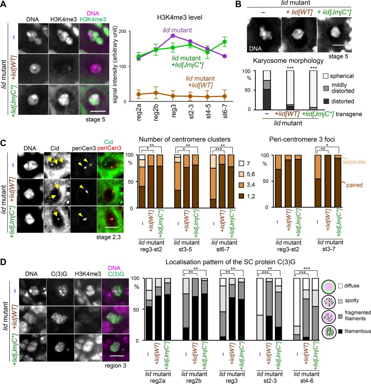
Fig 5. Kdm5/Lid demethylase activity is dispensable for meiotic chromatin reorganisation.
(A) Distribution and intensity of H3K4me3 on meiotic chromosomes in a Kdm5/lid mutant (lid10424/k06801) without a transgene (–), the Kdm5/lid mutant carrying a wild-type transgene (lid[WT]) and the Kdm5/lid mutant carrying a catalytically inactive transgene (lid[JmjC*]). Images of the karyosome in stage-5 oocytes were taken and the contrast has been enhanced using identical conditions. The total signal intensity of H3K4me3 on the karyosome was measured as described in Materials and Methods. Error bars indicate standard errors. n≥6. The signal intensity in the Kdm5/lid mutant carrying no transgenes (–) or the lid[JmjC*] transgene is significantly different from the one carrying the lid[WT] transgene at all stages (p<0.001). (B) Rescue of karyosome defects of the Kdm5/lid mutant by both the wild-type transgene (lid[WT]) and demethylase-inactive transgene (lid[JmjC*]). Karyosome morphology was observed in the Kdm5/lid mutant oocytes carrying no transgenes (–), the lid[WT] transgene or the lid[JmjC*] transgene, and was classified as in Fig 1C. *** indicates a significant difference in the pattern of distribution from the control (p<0.001). n≥18. (C) Centromere pairing and clustering do not require the demethylase activity of Kdm5/Lid. Centromeres highlighted by the Cid antibody are indicated by arrows, and foci of pericentromeric dodeca satellite specific to chromosome 3 (periCen3) are indicated by arrowheads. The numbers of centromere (Cid) clusters were counted for each group of stages (n≥12). Oocyte nuclei with tightly paired (<1 μm) or separate (≥1 μm) periCen3 foci were counted for each group of stages (n≥11). ***, ** and * indicate significant differences (p<0.001, p<0.01 and p<0.05, respectively). (D) SC morphology is not affected by loss of the Kdm5/Lid demethylase activity. Filamentous structures of C(3)G observed in region 3 oocytes from the Kdm5/lid mutant carrying the lid[WT] or lid[JmjC*] transgene, and a spotty appearance observed in the Kdm5/lid mutant alone. The SC component C(3)G and H3K4me3 were co-stained, imaged and contrast-enhanced using identical conditions. The morphology of the C(3)G-containing structure was categorised and counted for each group of stages (n≥10). ***, ** and * indicate significant differences (p<0.001, p<0.01 and p<0.05, respectively). Scale bars = 5 μm in all images.

