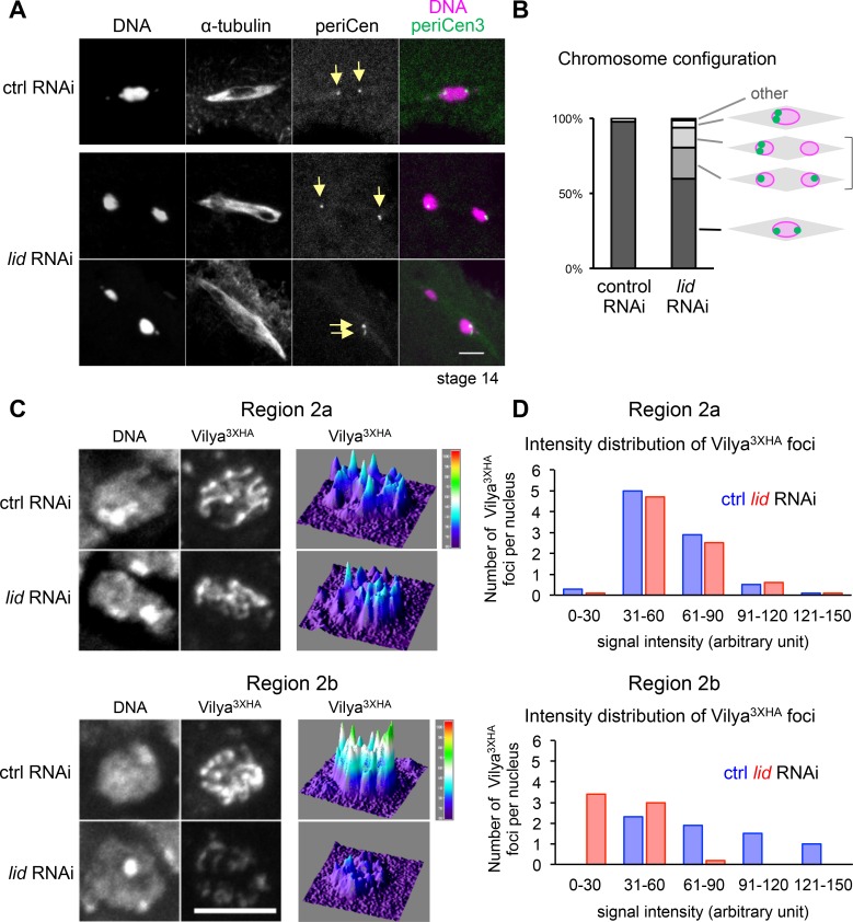
Fig 6. Abnormal chromosome positioning and orientation in prometa/metaphase I and a potential reduction in crossovers in oocytes lacking Kdm5/Lid.
(A) Mis-positioned chromosomes with normal spindle morphology in Kdm5/lid RNAi oocytes in prometa/metaphase I in comparison to control RNAi. Chromosome orientation was assessed by in situ hybridisation using dodeca satellite as a pericentromere 3 probe (arrows). Scale bar = 5 μm. (B) Quantification of chromosome configuration in control and Kdm5/lid RNAi oocytes. The “other” category includes a meiotic figure with more than two foci of the pericentromere 3 signal. The frequency of mis-oriented pericentromere 3 in Kdm5/lid RNAi is significantly different from the control RNAi (p<0.05). n≥45. (C) Localisation and signal intensity of Vilya-3xHA foci in meiotic nuclei in region 2a and 2b of control and Kdm5/lid ovaries expressing HA-tagged Vilya. Scale bar = 5 μm. (D) The signal intensity of Vilya3xHA foci in region 2a and 2b meiotic nuclei of control and Kdm5/lid RNAi ovaries expressing HA-tagged Vilya. The graphs show the numbers of foci per meiotic nucleus with the maximum signal intensity in indicated ranges. Foci with a signal intensity lower than 30 are significantly more frequent in Kdm5/lid RNAi than in control (p<0.001). Intensities of ≥66 Vilya3XHA foci have been measured for each region of each genotype.

