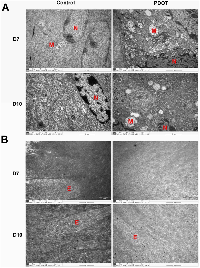
Fig 10. Electromicroscopic photographs of ameloblasts of mandibular first molar tooth germ in baby mice of postnatal day 7 and 10 from mothers treated with 4P- PDOT, a melatonin receptor blocker.
A, ultrastructure of ameloblasts, 13500x; B, hydroxyapatite matrix and enamel rod, 24500x. Labels in photos: E, enamel rod; M, mitochondria; N, nucleus. Arrows point to rough surfaced endoplasmic reticulum.

