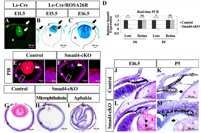
Fig 1. Varying degrees of microphthalmia or aphakia in Smad4-cKO mutants compared to the wild type mice.
(A) The expression pattern of Cre recombinase was specifically observed in the lens and cornea by detecting GFP fluorescence (arrows). (B-C) By crossing Le-Cre mice to ROSA26 reporter mice, Cre-mediated recombination was specifically observed in the lens, cornea and eyelids (arrows). (D) Real-time qPCR was employed to detect the expression of Smad4 in lens and retina at P0 and P8, respectively. n = 4, *P<0.05. (E-F) Immunostaining was performed to detect the expression of Smad4 in cKO eyes of E18.5, which showed the specific loss of Smad4 in the lens but not retina (arrows). (G-I) Microphthalmia and aphakia were observed in Smad4-cKO mutants at P7. In the aphakia mutant, the retina appeared pleated and corrugated in the center of the eye ball, while in microphthalmia, the retina formed a cup-structure and showed a proper orientation. (J-M) Abnormal development of the CMZ presented in the Smad4-cKO compared to the wild type mice at E16.5 and P5. Presence of cortical vacuoles in the mutant lens (arrowheads). At E16.5, the Smad4-cKO mice presented normal forward extension and early development of the ciliary body and iris as shown by thinning of the periphery of the retina (CMZ). But at P5, the iris stroma showed hypoplasia with the cornea, iris, and lens attached to each other (round frame). Furthermore, the neuroepithelium failed to fold backward to form ciliary body (arrows). E, embryonic; P, postnatal; M, month; L, lens; C, cornea; R, retina; I, iris; Ci, ciliary body; CMZ, ciliary marginal zone.

