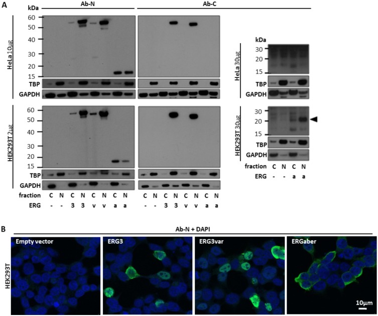
Fig 3. Analysis of subcellular localization of ERG isoforms by western blot and confocal microscopy.
(A) Protein lysates from HeLa and HEK293T cells (10μg and 2μg, respectively) transiently transfected by ERG3 (3), ERG3var (v) and ERGaber (a) in pcDNA3.1 vector or by empty vector (-) were analyzed by western blot to determine subcellular localization of individual ERG isoforms (left panel). ERG3 and ERG3var were detected dominantly in the nuclear fraction of protein lysate (N) in both cell lines, while ERGaberN was found in both cytoplasmic (C) and nuclear fractions in HeLa cells and dominantly in cytoplasmic fraction in HEK293T cells. ERGaberC was not detected in any cell line using these protein loads. Using higher load of protein lysate (30μg), sensitive visualization kit and longer exposition to X-ray films ERGaberC was detected in nuclear fraction of HEK293T but not HeLa cells (right panel, black arrow points to corresponding protein band). TBP and GAPDH proteins were used to control protein load and separation of cellular fractions. (B) HEK293T cells were transiently transfected by ERG3, ERG3var and ERGaber isoforms in pcDNA3.1 vector or by empty vector. Forty-eight hours after transfection the presence and the subcellular localization of ERG isoforms was analyzed by confocal microscopy using Ab-N antibody. Nuclei were stained by DAPI. The scale bar represents 10μm.

