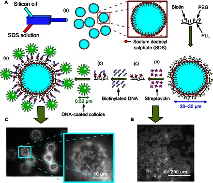
Fig. 1. DNA functionalization of oil droplets and colloids.

(A) Cartoon representing various stages of the sample preparation: (a) SDS-stabilized droplets are prepared by mixing 10 mM SDS and silicone oil in a microfluidic device; (b) PLL-PEG-biotin adsorbs at the SDS-stabilized ODs that are negatively charged because of the sulfate head group of the SDS surfactant; (c) Texas Red–labeled streptavidin linkers are then attached to the biotin heads on the ODs from the solution; (d) biotinylated ssDNA (A DNA) is then added, attaching to these streptavidin linkers; and (e) green fluorescent PS colloids coated with complementary A′ DNA are then allowed to bind via DNA hybridization. (B) Fluorescence image of the ODs after attaching the fluorescent streptavidin from solution. (C) Typical image showing the fluorescent colloids hybridized to the OD surfaces with a zoom onto the south pole of the droplet.
