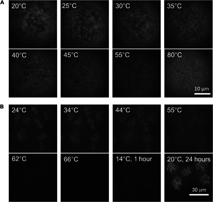
Fig. 3. Thermal recyclability of colloidal binding at the interface.

(A and B) Sequence of fluorescence images showing heating of a sample of 0.57-μm colloids grafted to ODs using colloids fully covered with A′ DNA (~1.5 mM SDS and 50 mM NaCl concentrations) (A) and where three-fourths of the A′ DNA is replaced by nonbinding B (~2.5 mM SDS and 50 mM SDS) (B). In all images, we focus on the south pole of the ODs using the same illumination and exposure time. (A) For full coverage, most colloids remained on the OD surface even when heating to 80°C, although they became increasingly mobile, which is expressed in the increasingly blurry appearance of the images. (B) For samples with reduced number of binding sites on the colloids, melting was set at ~56°C, and at ~70°C, almost all colloids have come off the ODs. The image taken shortly after cooling the sample (over a period of 1 hour) to 14°C showed only a few colloids bound to the ODs. The same sample recovered similar coverage and aggregation state as the initial samples after 1 day (right-most bottom image).
