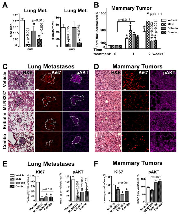
Figure 3. MLN8237 and MLN8237/eribulin combination reduces metastases in vivo.
A Quantification of size and number of pulmonary metastases as in (C). B Quantification of mammary tumor growth in MDA-MB-231LN-xenografts based on bioluminescence imaging (average radiance, p/s/cm2/sr) normalized to initial tumor volume (before drug application) at time point zero, referenced as 100%; one-way ANOVA: vehicle vs. treatments at each time point. C–D Representative images of H&E and F-IHC staining of pulmonary tissue (metastases defined by the white outline based on H&E), or D Mammary MDA-MB-231LN-xenograft tumor with anti-Ki67/red, -pAKT/purple antibodies. Scale bar - 30μm E–F Quantification of F-IHC as in (C–D); multiple metastases/or tumors; 5–6 animals, n=3; % to control; one-way ANOVA: vehicle vs. treatments;

