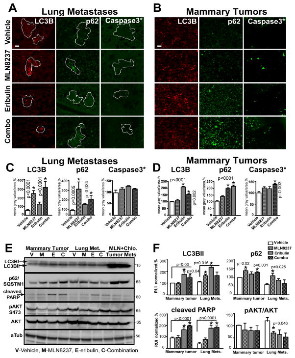
Figure 4. MLN8237/eribulin combination induce cytotoxic autophagy and apoptosis.
A–B Representative images of F-IHC staining of pulmonary tissue (A-metastases defined by the white outline) or B mammary MDA-MB-231LN-xenograft tumor with -LC3B/red, -SQSTM1/p62/green, -cleaved-caspase-3*/green antibodies. Scale bar - 30μm C–D Quantification of F-IHC as in (A–B); multiple metastases/or tumors; 5–6 animals, n=3; % to control; one-way ANOVA: vehicle vs. treatments. E WB analysis of LC3B, p62, cleaved PARP, pAKT, AKT, alpha-tubulin (loading control) with respective antibodies in MDA-MB-231LN cells isolated from vehicle-treated mice, expanded in vitro and treated with drugs for 48h as indicated; last two lanes contain lysates treated with MLN8237+chloroquine (Chlo.). F Quantification of digital images as in (C) using GeneTools software, mean grey value of corresponding band/area (% of vehicle, normalized to tubulin) ±S.E.M, n=3, one-way ANOVA: vehicle vs. treatments.

