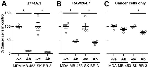
Figure 1.
Macrophages reduce breast cancer cell viability in the presence of trastuzumab. J774A.1 (A) or RAW264.7 (B) macrophages were plated in 48 well plates with MDA-MB-453 or SK-BR-3 breast cancer cells at a 4:1 effector:target cell ratio (2.5×10⁴:6.25×10³ cells) and 1 μg/ml trastuzumab (Ab) or PBS vehicle (-ve) was added 24 hours later. Following 72 hours, cells were harvested and the remaining number of cancer cells quantitated by flow cytometry. The number of live cancer cells in each sample is shown as a fraction of the corresponding vehicle control. C, cell numbers following incubation of cancer cells as in A,B but without macrophages. Error bars represent standard errors. Student's t-test was performed to indicate statistical significance (denoted by *; p < 0.05).

