Figure 3.
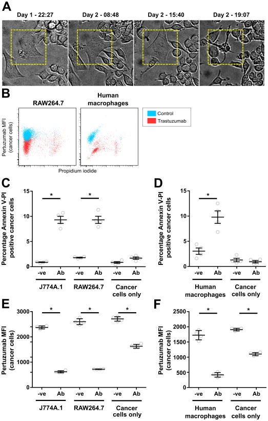
RAW264.7, J774A.1 and human monocyte-derived macrophages induce similar levels of apoptosis in opsonized cancer cells. Macrophages and cancer cells were plated at an effector:target cell ratio of 4:1 in a T25 culture flask (3×10⁶:7.5×10⁵ cells), 1 μg/ml trastuzumab was added 18-24 hours later and the cells were imaged. A, individual frames showing an SK-BR-3 cell undergoing cell death in a long-term light microscopy imaging experiment for a co-culture of SK-BR-3 and RAW264.7 cells in the presence of 1 μg/ml trastuzumab. Times of acquisition of each image are indicated. B,C,D, MDA-MB-453 cells were plated alone or co-incubated with RAW264.7, J774A.1 or human monocyte-derived macrophages at a 4:1 effector:target cell ratio in the presence of 1 μg/ml trastuzumab (Ab) or PBS vehicle (-ve) for 36 hours. B, representative dot-plots for pertuzumab fluorescence vs. PI fluoresence for cancer cells from co-cultures of RAW264.7 or human macrophages with cancer cells in the presence (trastuzumab) and absence of antibody (control). C,D, fraction of annexin V, PI double-positive cancer cells in co-cultures determined by flow cytometry. E,F, samples shown in C and D, respectively, were stained with fluorescently labeled pertuzumab after harvesting and the mean fluorescent intensity (MFI) of pertuzumab on the cancer cell populations determined. Error bars represent standard errors. Student's t-test was performed to indicate statistical significance (denoted by *; p < 0.05).

