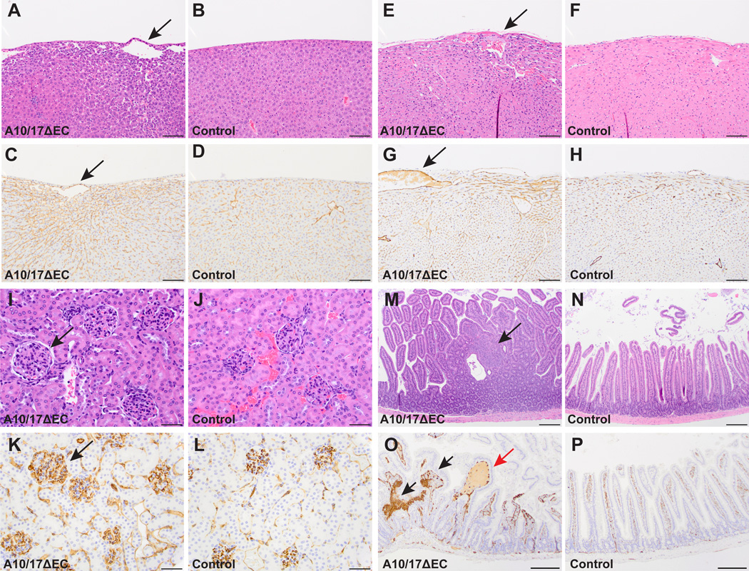
Figure 2. A10/17ΔEC mice exhibit defects in organ-specific vascular beds.
Histopathological analysis of hematoxylin and eosin (H&E) or CD31 (endothelial cell marker) stained specimens reveal defects in vascular beds of the liver, heart, kidney, and small intestine of A10/17ΔEC mice. A to D, H&E-stained A10/17ΔEC liver (A) shows enlarged vessels (arrow) near the liver surface not present in controls (B). The enlarged vessels are CD31+ (C, arrow) and are associated with changes in surrounding sinusoidal endothelium not seen in controls (D). E to H, an A10/17ΔEC heart (E) shows an enlarged subepicardial vessel and myocardial hypercellularity compared to a control (F). The enlarged vessel in the A10/17ΔEC heart (G, arrow) is CD31+ and myocardial hypercellularity is associated with increased CD31 staining compared to the control (H). I to L, an H&E stained A10/17ΔEC kidney shows larger, more hypercellular glomeruli (I, arrow) than a control (J). CD31 staining is increased in A10/17ΔEC glomeruli (K, arrow) compared to control glomeruli (L). M to P, small intestine of an A10/17ΔEC mouse contains hyperplastic polyps (M, arrow) not present in controls (N). Polyps contain abnormal nests of CD31+ cells (O, black arrows) and are occasionally fluid-filled (red arrow) in contrast to the regular architecture of control villi (P). H&E micrographs shown are representative of 8-week old animals analyzed for each genotype (n=3 A10/17ΔEC, n=3 A10flox/flox/A17flox/flox). See supplemental Figure II for quantification. Scale bars, 100µm (A-H), 50µm (I-L), and 200µm (M-P).

