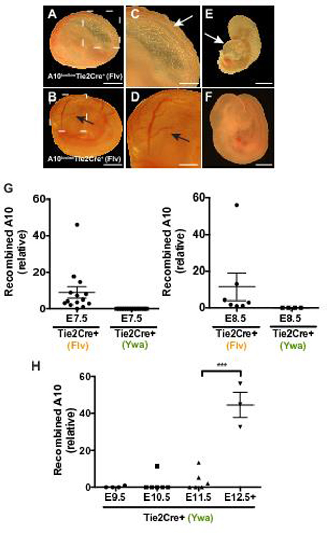
Figure 4. Deletion of ADAM10 in endothelial cells using a different Tie2-Cre transgenic driver results in abnormal embryonic development and earlier Cre-mediated recombination.

A to F, Representative images of whole mount preparations of embryos and yolk sacs harvested at E10.5. A to D, a yolk sac isolated from an E10.5 A10flox/flox-Tie2-Cre+(Flv) embryo appeared wrinkled (A,C) and devoid of the large vessels seen in the yolk sac of the A10flox/wt-Tie2-Cre+(Flv) littermate control (B,D, arrows). The A10flox/flox-Tie2-Cre+(Flv) embryo is smaller (E) than the normally developing littermate control (F). A10flox/flox-Tie2-Cre+(Flv) embryos had an enlarged pericardial sac (E, arrow), and were dead as determined by the absence of a heartbeat (11 out of 12 embryos). G, Total DNA from E7.5-E8.5 Tie2-Cre+(Flv) embryos or from E7.5-E8.5 Tie2-Cre+(Ywa) embryos were subjected to qPCR to determine levels of recombined Adam10 in embryos. 13 out of 14 E7.5 Tie2-Cre+(Flv) embryos (n=6 A10flox/wtTie2-Cre+(Flv); n=8, A10flox/floxTie2-Cre+(Flv)) showed detectable recombined Adam10 product while no recombined Adam10 was detected in E7.5 Tie2-Cre+(Ywa) embryos (n=7, A10flox/floxTie2-Cre+(Ywa)). 7 out of 7 E8.5 Tie2-Cre+(Flv) embryos (n=6 A10flox/wt Tie2-Cre+(Flv); n=1, A10flox/flox Tie2-Cre+(Flv)) had detectable recombined Adam10 while no recombined Adam10 was detected in 4 A10flox/flox Tie2-Cre+(Ywa) embryos. Samples with no detectable signal are plotted as zero. H, Total DNA prepared from A10flox/floxTie2-Cre+(Ywa) embryos between E9.5 and E13.5 were analyzed as described for G by qPCR. Recombined Adam10 was detected in 1 of 4 E9.5 embryos, 1 of 6 E10.5 embryos, and 3 of 6 E11.5 embryos. Recombined Adam10 was detectable in all embryos between E12.5 and E13.5 (n=3). Expression levels in G and H were normalized to Gapdh or the promoter region of Mrc1. Data shown as mean ± SEM. *** signifies p<0.001 in two-tailed student’s t-test. Scale bars, 1mm (A,B,E,F), 500µm (C,D).
