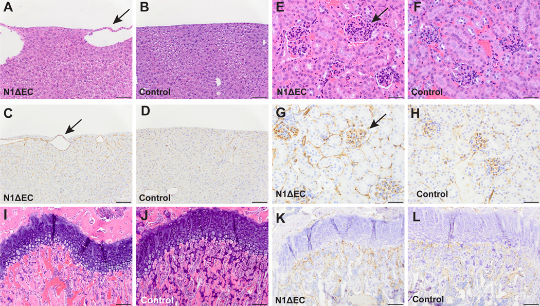
Figure 5. Several organ-specific vascular beds are affected in N1ΔEC mice.
A to L, Histopathological analysis of H&E- and CD31-stained specimen from N1ΔEC mice revealed defects in vascular beds of the liver, kidney, and femur. A to D, the livers of N1ΔEC mice contain enlarged surface vessels (A, arrow) not present in controls (B). The enlarged vessels are CD31+ (C, arrow) and associated with mild changes in nearby sinusoidal endothelium not seen in the control (D). E to H, glomeruli of an N1ΔEC mouse (E, arrow) are larger than control glomeruli (F). N1ΔEC glomeruli show increased CD31 staining (G, arrow) compared to controls (H). Abnormally enlarged vessels are seen under the femoral growth plate of an N1ΔEC mouse (I, arrow) in contrast to the small vessels seen in a control mouse (J, arrow). The enlarged vessels in N1ΔEC femurs are CD31+ (K) and are immediately adjacent to growth plate, and are smaller in control mice (L, see supplemental figure VI for higher magnification images and quantification). H&E micrographs are representative of 8-week old animals analyzed for each genotype (n=4 N1ΔEC, n=4 N1flox/flox), see supplemental Figure XX for quantification. Scale bars, 100µm (A-D, I-L), 50µm (E-H).

