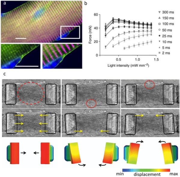
Figure 2. Control of cell contractile forces using optogenetics.
(a) Skeletal muscle fiber isolated from mice expressing EYFP-tagged channelrhodopsin-2 (green), which localizes to the cell membrane, including the T-tubule membrane invaginations that surround sarcomeric alpha-actinin (magenta). Scale bar shows 10 µm. (b) Explanted soleus muscles contracted in response to blue light with dependence on light pulse duration and intensity. “Functional expression of ChR2 in skeletal muscle.” by Bruegmann et al. [45], licensed under CC BY 4.0 (c) Devices comprised of skeletal myotubules expressing channelrhodopsin-2 in matrigel-collagen gels surrounding PDMS cantilevers can be induced to contract axially or rotationally by controlling the region of illumination (circled in red). Reproduced with permission from Sakar et al.[46]

