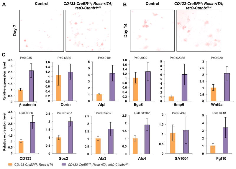
Figure 2. Upregulated expression of dermal papilla signature genes in ΔN-β-catenin-expressing CD133+ DP cells in hydrogel culture.
A. AP staining of cultured spheroids formed by control (left) and ΔN-β-catenin-expressing (right) CD133+ DP cells in hydrogel at day 7. B. AP staining of cultured spheroids formed by control (left) and ΔN-β-catenin-expressing (right) CD133+ DP cells in hydrogel at day 14. C. Quantitative real-time PCR analysis of expression of DP signature genes, including β-catenin (Ctnnb1), Corin, CD133, AP (Alpl), Integrin α8 (Itga8), Sox2, Bmp6, Wnt5a, Alx3, Alx4, S100A4, Fgf10. The Y-axis represents fold change in expression with the level in control set to 1 (n=6).

