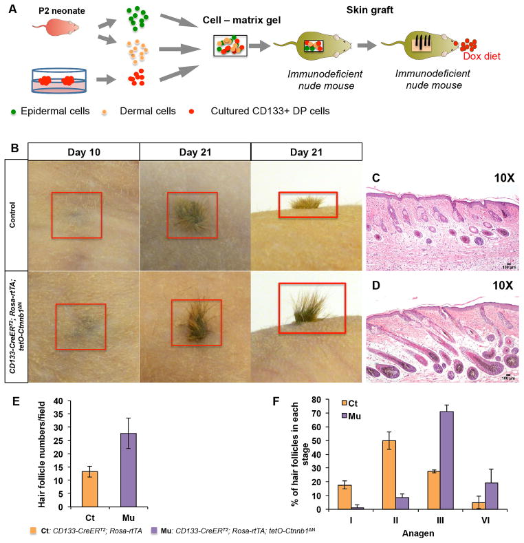
Figure 3. ΔN-β-catenin-expressing CD133+ DP cells induce accelerated hair growth in reconstituted skin.
A. Schematic representation of hair reconstitution assays. Spheroids were released by disaggregating hydrogels and dissociated to release CD133+ DP cells. Same number of control or β-catenin-expressing CD133+ DP cells mixed with P2 dermal cells and epidermal cells was grafted onto nude mice and observed for hair follicle formation, respectively. B. Appearance of newly formed hairs at day 10 and day 21 after grafting. Upper panels: control group containing normal CD133+ DP cells; lower panels: CD133-CreERT2; Rosa-rtTA; tetO-Ctnnb1ΔN group containing β-catenin-expressing CD133+ DP cells (n=5). Skin biopsies from hair bearing wound area of mice grafted with CD133-CreERT2; Rosa-rtTA; tetO-Ctnnb1ΔN DP cells (D) or control CD133+ DP cells (C) were stained with H&E and photographed at indicated stages. Scale bars: 100 μm. E. Hair follicle numbers in reconstituted skin formed by either control and β-catenin-expressing CD133+ DP cells were counted in each field after H&E staining. At least 3 random fields were selected and counted for each reconstituted skin sample (n=3). F. Hair follicles at different anagen stages were counted on H&E stained reconstituted skin samples according to the classification system published previously (1). At least 3 random fields were picked and counted for each reconstituted skin sample (n=3).

