
Fig. 1.
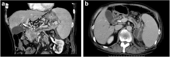
Abdominal CT showing a low-density mass, measuring 40 × 30 mm, in the pancreatic head associated with a marked dilatation of the bile duct and distal pancreatic duct (a). There was also an irregular-shaped cystic lesion spreading from the pancreatic tail to the splenic hilum, suggestive of a pancreatic pseudocyst associated with severe obstructive pancreatitis (b)