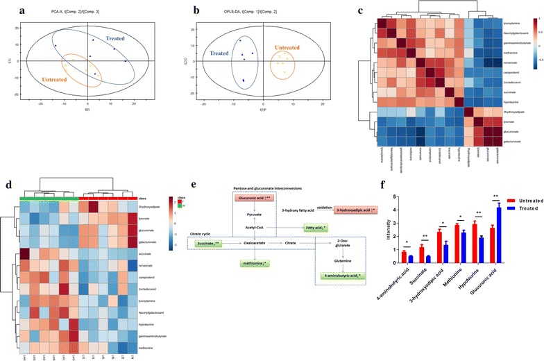
Fig. 4.

The metabolic profiles in liver tissue. The HFD induced hyperlipidemic rats were treated with or without BC for 4-week, liver tissues were collected, and metabolomics analysis was made by GC/MS. a PCA score plots of liver samples from BC treated group and untreated group; b scores plots of OPLS-DA between untreated group and BC treated group; c Pearson’s correlations of the quantities of the 13 metabolites determined from rat liver samples; d heat map showing the FC of 19 metabolites. Shades of green represent FC decrease while red represent FC increase; e simplified draft illustrating perturbed pathways involved; f differential metabolites between groups in the liver. Values were showed as mean peak intensities ± SEM. *P < 0.05; **P < 0.01, compared with untreated (HFD) group
