
Figure 1.
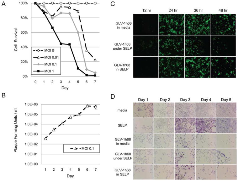
GLV-1h68 infects and lyses 8505c anaplastic thyroid cancer cells equally well in silk-elastin-like protein polymer (SELP) as in solution. (A) To assess the sensitivity of 8505c cells to GLV-1h68, virus at varying multiplicities of infection was added to cells in media and cell viability measured daily for 1 week by lactate dehydrogenase (LDH) assay. (B) Viral proliferation was assessed by measuring daily viral titers from the supernatants of the multiplicity of infection (MOI) 0.1 wells. (C) The ability of GLV-1h68 at MOI 1 to infect a monolayer of 8505c cells was assessed by green fluorescent protein (GFP) expression between (A) GLV-1h68 in media versus (B) GLV-1h68 in media under a solidified layer of SELP gel versus (C) GLV-1h68 in a matrix of SELP gel. (D) The same groups were used to assess cytotoxicity by assessing cell viability over 5 days using crystal violet staining. [Color figure can be viewed in the online issue, which is available at wileyonlinelibrary.com.]