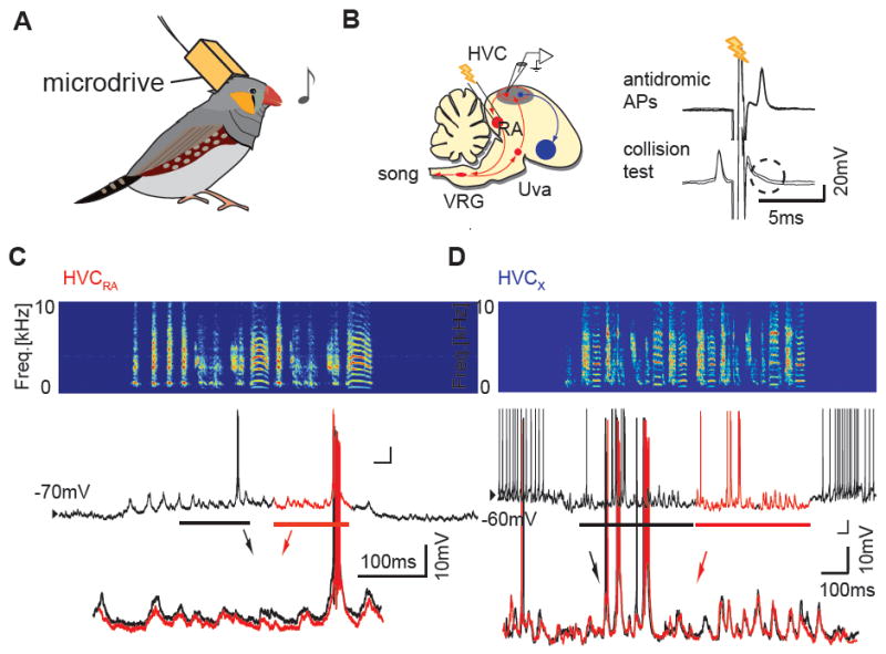
Figure 7. Examples of intracellular recordings of HVC PNs in singing birds.

A, Schematic of sharp intracellular recordings in singing birds. B, Left: Schematic diagram of song motor pathway (red) and a part of the basal ganglia pathway (blue; Area X). Right: An example of antidromic identification of an HVCRA neuron using a spike collision test. C, An example of synaptic and action potential activity of an identified HVCRA neuron. Top: sonogram, Middle: membrane potential, Bottom: enlarged and overlaid membrane potential traces from two consecutive motifs reveal highly stereotyped synaptic activity. D, An example of synaptic and action potential activity of a putative HVCX neuron recorded in another bird; overlaid membrane potential traces from two consecutive motifs also reveal stereotyped synaptic activity.
