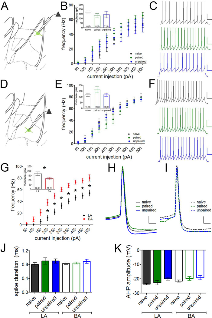
Figure 7. Fear memory encoding does not affect PV-IN intrinsic excitability.
Action potentials were elicited in PV-INs in LA (A–C) and BA (D–F) by somatic current injections of increasing amplitude. A. Recording configuration in B–C. No differences in intrinsic excitability were observed among groups in LA PV-INs (B, representative traces at the 200 pA current injection in C). D. Recording configuration in E–F. No differences in intrinsic excitability were observed among groups in BA PV-INs (E, representative traces at the 200 pA current injection in F). Scales = 50 pA × 25 ms. G. In naïve animals, PV-INs in LA were less excitable than PV-INs in BA, requiring more current to fire a single action potential (rheobase current; inset) and firing fewer action potentials to current injections greater than 150 pA. H–K. Individual spike characteristics were quantified in LA (H) and BA (I) at rheobase. Overlays of representative traces are shown. Scales = 10 pA × 2 ms. No differences were observed among groups or between nuclei in any spike parameter, including spike duration (J) and after hyperpolarization (AHP) amplitude (K). n/group indicated on graphs. G, repeated-measures ANOVA following by Holm-Bonferroni or independent samples t-test (inset). * p < 0.05. Data presented as mean ± SEM.

