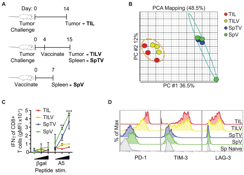
FIGURE 2.
Genome-wide mRNA expression of tumor-specific CD8+ T cells segregates into two functionally distinct clusters. (A) Schematic of the origin of tumor antigen-specific T cells is shown. AH1-specific CD8+ T cells were tetramer-sorted from CT26 tumors of mice that had (TILV) or had not (TIL) been vaccinated, and spleens of vaccinated mice that either harbored (SpTV) or did not harbor tumors (SpV). (B) Messenger RNA from the groups described in A was interrogated by microarray. Principal component analysis (PCA) divided genome-wide mRNA expression of the indicated cell populations into two distinct clusters, tumor-specific CD8+ T cells from the spleen or the tumor. Five to ten mice were pooled per group, and each group was repeated 3 times. (C) AH1-specific CD8+ T cells described in A were stimulated ex vivo with increasing concentrations of the indicated peptide and assayed for IFNγ production. (D) AH1-specific CD8+ T cells described in A were analyzed for inhibitory receptor expression. The grey histogram represents naive CD8+ T cells from a spleen as a negative control for inhibitory receptor expression. C and D are representative data of at least 2 independent experiments, n=1-4 biological replicates, and error bars=SD.

