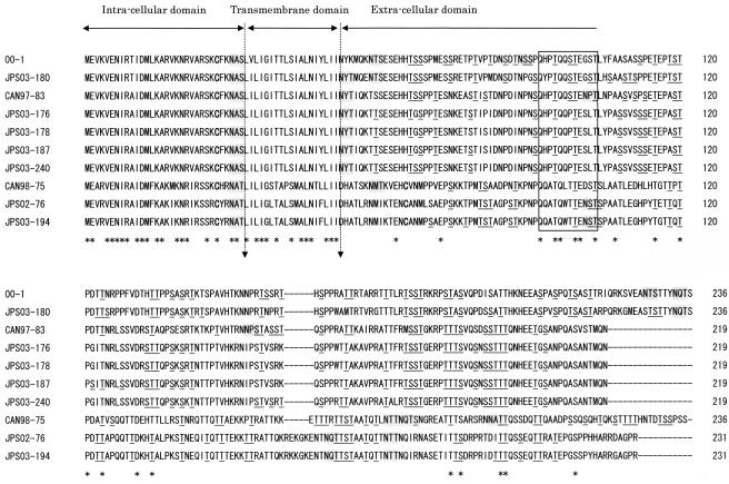
FIG. 3.
Comparison of the predicted amino acid sequences of G of hMPV isolates. The predicted amino acid sequence of G of hMPV with cysteine residues is shown in boldface type, potential N-linked glycosylation sites are shaded in gray, and potential O-glycosylation sites are underlined. Dashes indicate gaps introduced to maximize the alignment or to denote the absence of corresponding amino acids. The asterisks underneath each alignment denote amino acid identity among all sequences. Proposed intracellular, transmembrane, and extracellular domains are indicated above the sequences. The square indicates the positions of conserved amino acid residues.

