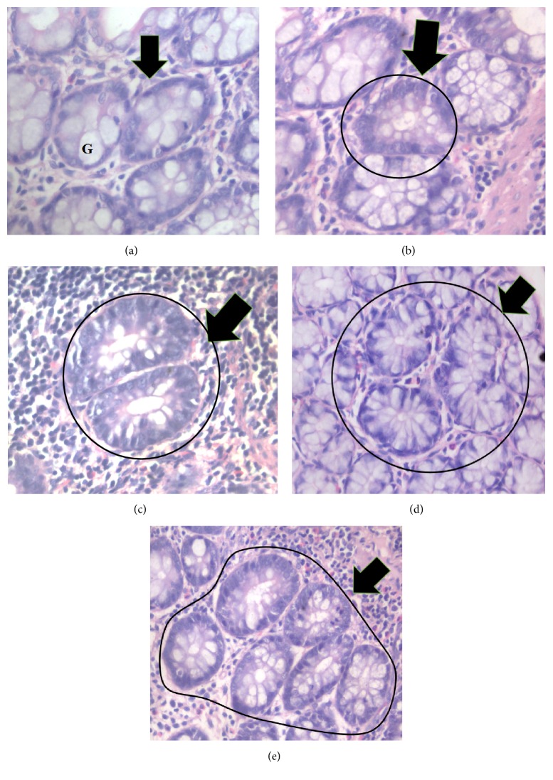
Figure 2.
Photomicrograph of (a) normal crypt, (b) 1 crypt, (c) 2 crypts, (d) 3 crypts, and (e) ≥4 crypts stained with hematoxylin and eosin. Normal crypt is characterized by constant internal and diameter between the crypts and higher proportion of goblet cells (G), while ACF exhibit darker stain with reduced number of goblet cells (400x magnification).

