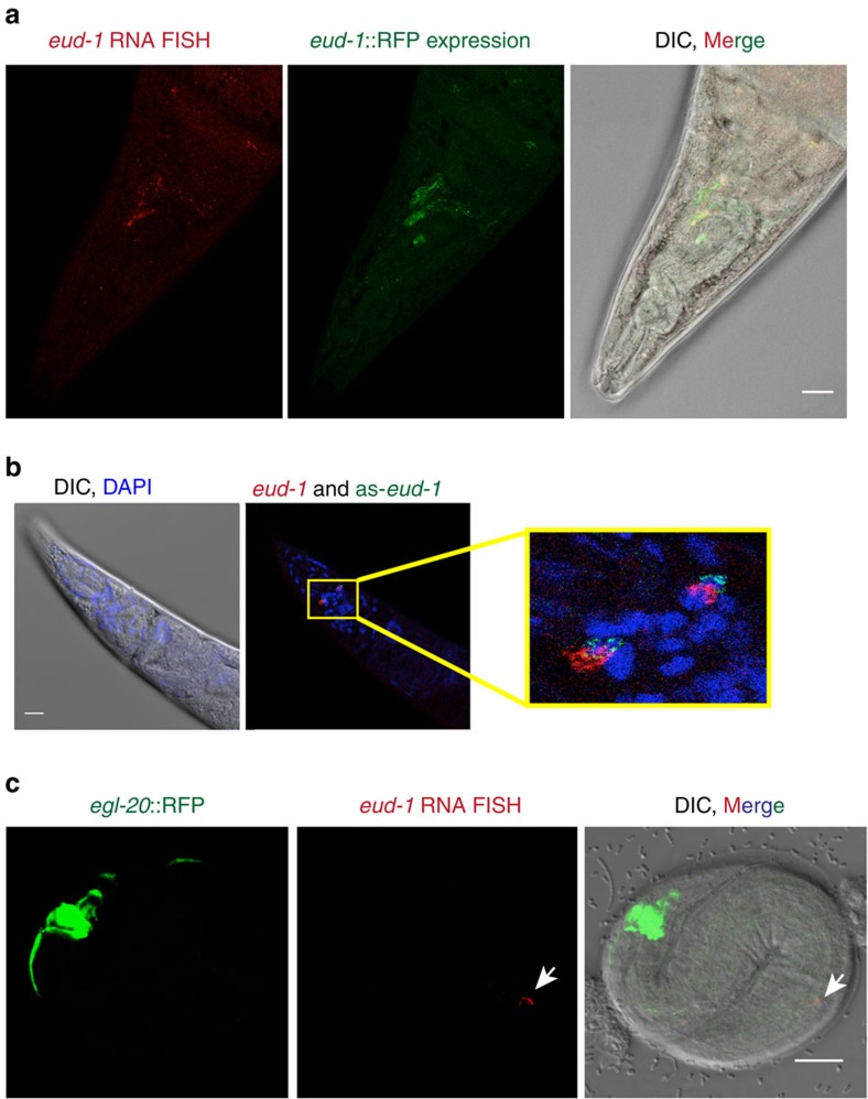
Figure 4. Fluorescent in situ hybridization (FISH) of eud-1 and as-eud-1 reveals co-expression of both transcripts.
FISH probes were designed as described in the Methods section. Photographs in a and b show adult animals, photographs in c show a J1 stage larvae, which in P. pacificus is still in the egg shell. (a) eud-1 FISH (red, left image) and an eud-1::RFP reporter construct (green, central image) show the same expression pattern in several head neurons. The image at the right represents a merger of both and differential interference contrast (DIC) microscopy. Note that not all eud-1-expressing cells are visible in this plane of focus. (b) Head area of an adult worm with DIC and 4,6-diamidino-2-phenylindole staining (left image) and co-expression of eud-1 (red) and as-eud-1 (green) as revealed by FISH probes. Both transcripts are expressed at multiple foci, two of which are shown in this plane of focus (inset). Overlapping fluorescence (yellow) was seen in all animals observed, but not in all cells. The expression pattern was highly consistent among multiple adults (n>20). See Supplementary Movie for additional details of the partially overlapping expression of both transcripts. (c) Transgenic animals carrying an eud-1::as-eud-1 construct show eud-1 expression in head neurons already in the J1 stage, which is never seen in wild-type animals. egl-20::RFP (green, left image) is used as transformation marker. The same eud-1 FISH probe (red, central image) was used as above. The image at the right represents a merger and DIC microscopy. Supplementary Fig. 4 provides additional photographs for a and b. Scale bars, 10 μm.

