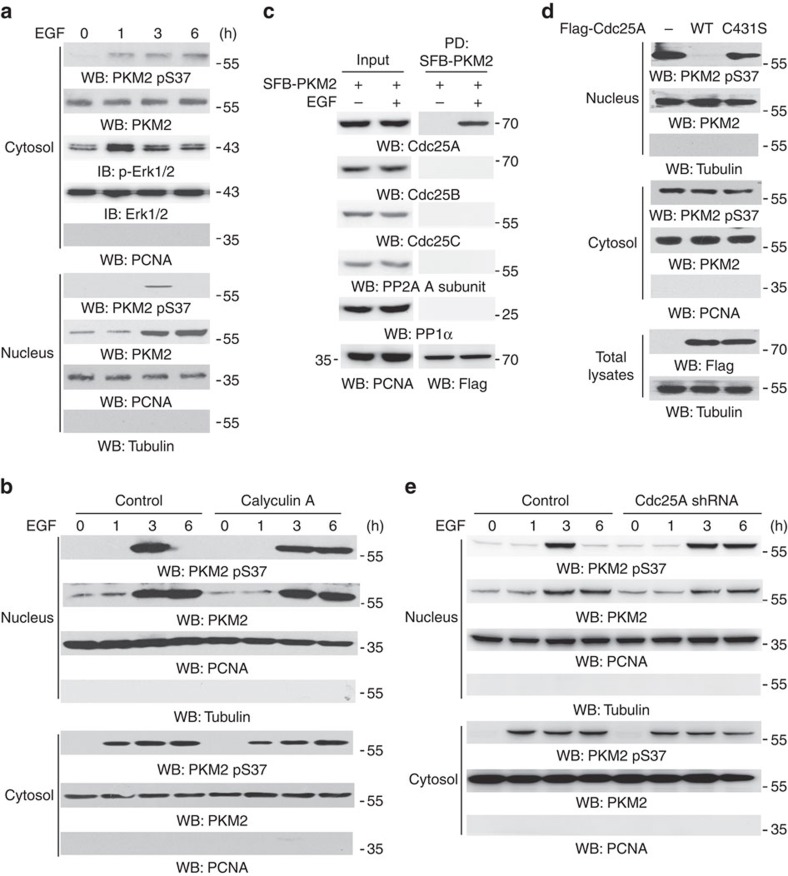
Figure 1. Nuclear PKM2 pS37 is dephosphorylated by Cdc25A.
Immunoprecipitation and immunoblotting analyses were performed with the indicated antibodies. Data are representative of at least three independent experiments. (a) U87/EGFR cells were treated with or without EGF (100 ng ml−1) for the indicated period of time. Cytosolic and nuclear fractions of the cells were prepared. (b) U87/EGFR cells were pretreated with calyculin A (25 nM) for 30 min before EGF (100 ng ml−1) treatment for the indicated period of time. Cytosolic and nuclear fractions of the cells were prepared. (c) U87/EGFR cells stably expressing SFB-PKM2 were treated with or without EGF (100 ng ml−1) for 4 h. Nuclear lysates were prepared and followed by a pull-down assay of SFB-PKM2 with streptavidin-agarose beads. PD, pull-down. (d) U87/EGFR cells were infected with or without a lentivirus expressing Flag-Cdc25A WT or a catalytically inactive Cdc25A mutant (Cdc25A C431S) and were treated with EGF (100 ng ml−1) for 3 h. Cytosolic and nuclear fractions of the cells were extracted. (e) U87/EGFR cells expressing a control shRNA or shRNA against Cdc25A were treated with EGF (100 ng ml−1) for the indicated period of time. Cytosolic and nuclear fractions of the cells were extracted.

