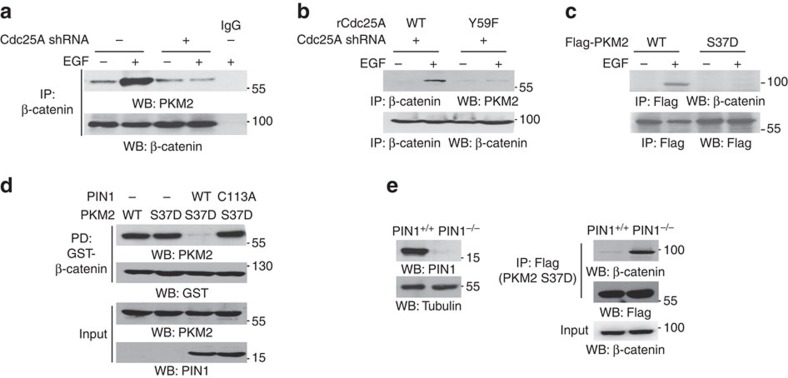
Figure 4. The binding of PKM2 to β-catenin requires PKM2 dephosphorylation.
Immunoprecipitation and immunoblotting analyses were performed with the indicated antibodies. Data are representative of at least three independent experiments. (a,b) U87/EGFR cells with or without depleted Cdc25A (a) and reconstituted expression of rCdc25A WT or rCdc25A Y59F (b) were treated with or without EGF (100 ng ml−1) for 6 h. (c) U87/EGFR cells infected with a lentivirus expressing Flag-PKM2 WT or PKM2 S37D were treated with or without EGF (100 ng ml−1) for 6 h. (d) Purified recombinant His-PKM2 S37D was incubated with or without purified WT PIN1 or PIN1 C113A for cis–trans isomerization assay. Meanwhile, an in vitro kinase assay was performed by mixing immobilized GST-β-catenin and active c-Src. After the reactions, pulled down GST-β-catenin was washed with PBS three times and incubated with His-PKM2 S37D overnight at 4 °C, followed by a pull-down assay of GST-β-catenin with glutathione agarose beads. (e) PIN1 WT (PIN1+/+) or PIN1 knockout (PIN1−/−) cells were infected with lentivirus expressing Flag-PKM2 S37D. Immunoprecipitated Flag-PKM2 S37D from both cells was washed with PBS for three times and subsequently incubated with nuclear lysates of U87/EGFR cells treated with EGF for 4 h.

