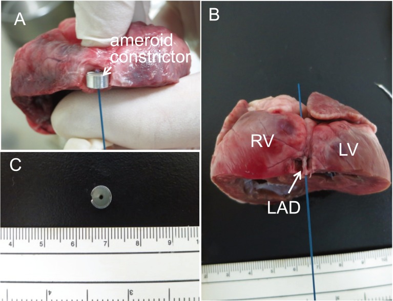
Fig. 4.
Photos of excised heart. (A) The heart was cut just distal to the mounting position of ameroid constrictor. A guidewire in a diameter of 0.035 inch was placed in the left anterior descending coronary artery, which was mounted by ameroid constrictor. (B) A guidewire in a diameter of 0.035 inch was placed in the left anterior descending coronary artery. The passage of the guidewire indicates the patency of the left anterior descending coronary artery. (C) Lumen of ameroid constrictor was not narrowed. RV: right ventricle; LV: left ventricle; and LAD: left anterior descending artery.

