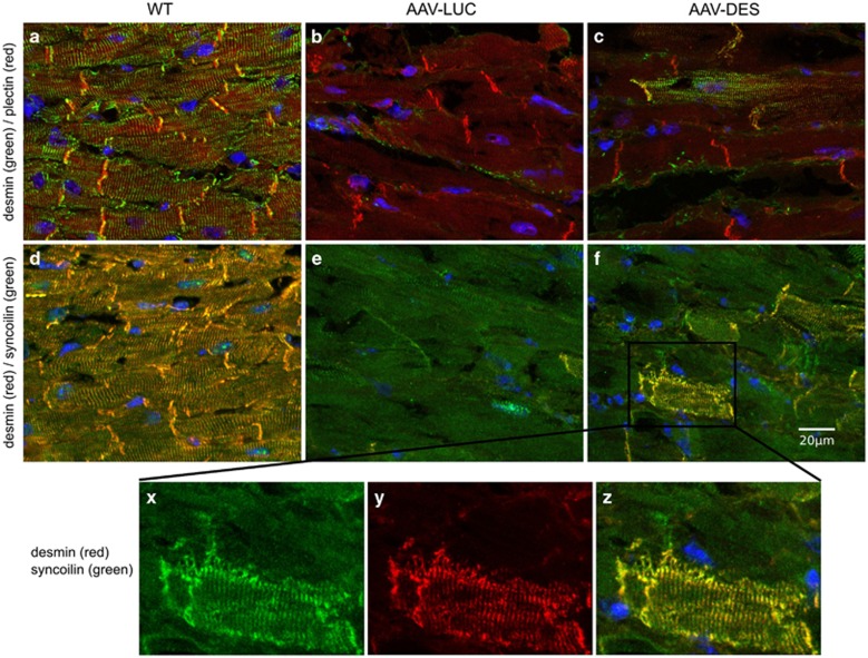
Figure 2.
Desmin/plectin/syncoilin co-staining with 4',6-Diamidin-2-phenylindol (DAPI). First column: (a, d) wild type (n=3). Second column: (b, e) AAV-LUC-treated desmin-deficient mouse (n=4). Third column: (c, f) and detail of (f) (x) syncoilin, y desmin, z merge) AAV-DES-treated desmin-deficient mouse (n=4). First row (a–c): desmin green, plectin red. Second row and details of (f) (d–f, x–z): desmin red, syncoilin green. While the subcellular distribution of plectin seems unaffected by the absence of desmin, the re-expression of desmin has a dramatic effect on the syncoilin staining pattern. Note that the AAV-DES treatment leads to the de novo formation of a cross-striated desmin–syncoilin staining pattern in a subset of DES-KO cardiomyocytes (x–z).

