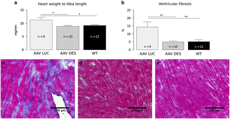
Figure 3.
Morphometric and histological analysis of hearts from mice treated with AAV-DES compared with AAV-LUC and wild-type controls. (a) Heart weight to tibia length ratios are significantly decreased in AAV-DES-treated animals (n=10) when compared with AAV-LUC controls (n=9) or wild-type mice (n=12). (b) Ventricular fibrosis is also significantly decreased in AAV-DES (n=10) and wild type (n=12) when compared with AAV-LUC (n=10). (c–e) Representative Masson's trichrome stainings of the left ventricle of AAV-LUC (n=10) (c), AAV-DES (n=10) (d) and wild type (n=12) (e). Note the intense fibrotic area (blue) depicted in (c). *P<0.05, **P<0.01.

