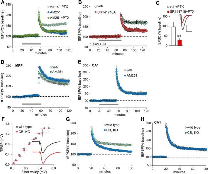
Figure 1.
LPP-LTP is endocannabinoid and CB1 dependent. A, The CB1 inverse agonist AM251 (5 µm, at horizontal bar) disrupts LPP-LTP with or without PTX (10 µm) in the ACSF bath. Veh-treated slices included those tested with (+) and without (−) PTX present. Potentiation, assessed 55–60 min after high-frequency stimulation, was greatly reduced in both AM251 groups (p < 0.0004, −PTX; p < 0.001, +PTX; n = 5 each) relative to veh. B, Perfusion of the CB1 antagonist SR141716A (5 µm) in the presence of 100 µm PTX significantly attenuated LPP-LTP, as assessed with field recordings (p = 0.0004, two-tailed t test for percentage of LTP in veh vs SR141716A). C, CB1 antagonist SR141716A also significantly reduced the percentage of LPP potentiation as assessed 25 min postinduction using voltage-clamp recordings of granule cells in slices treated with veh (n = 8) or 25 µm SR141716A (n = 6; **p < 0.005, two-tailed t test vs veh+PTX): traces show EPSCs collected before and 30 min after induction for veh slices. Calibration: 100 pA, 50 ms. D, E, AM251 did not reduce LTP of the MPP (D) or the S-C afferents of field CA1 (E; n = 5/group). F, Relationship of the peak amplitude of the fiber volley to the fEPSP slope (input/output curves) in the outer molecular layer of the DG with LPP stimulation in WT and CB1 KO mice: traces are averaged group responses after normalization of mean fEPSPs for individual slices to their peak amplitude. Group fEPSPs for WT and CB1 KO slices were indistinguishable. G, LTP in the LPP was severely impaired in CB1 KO mice (p < 0.0001; n = 10/group). H, LTP did not differ between CB1 KOs and WTs in field CA1 (p = 0.89, t(10) = 0.16: n = 6/group). All panels show results from field recordings (fEPSPs), except for the bar graph in C, which shows the results of whole-cell, voltage-clamp recordings.

