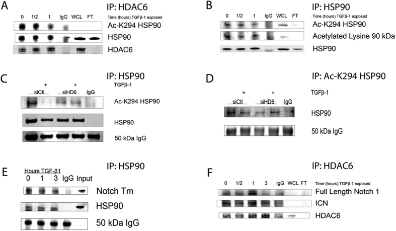
Figure 6. TGF-β1-induced deacetylation of HSP90 by HDAC6 in A549 cells.
(A,B,E,F) Immunoprecipitation of respective target antigens from whole cell lysates of serum-starved A549 cells treated with TGF-β1 (2.5 ng/ml) for the indicated time and examined by western analysis. (A) Western analysis for acetylated-K294 HSP90 co-precipitated with anti-HDAC6. (B) Western analysis of acetylated-K294 HSP90 immunoprecipitated with anti-HSP90. (C,D) Serum-starved A549 cells transiently transfected with siRNA targeting HDAC6, or non-specific siRNA treated with TGF-β1 for one hour. Cell lysates were immunoprecipitated with anti-HSP90 (C) or anti-Ac-K294 HSP90 (D) and western analysis of Ac-K294 HSP90 (C) or total HSP90 (D). (E,F) Immunoprecipitation of HDAC6 and HSP90, respectively, of same cell lysates as in (A,B) and examined by western analysis for co-precipitated full length Notch1 or transmembrane Notch1 (Notch Tm). WCL = whole cell lysate; FT = flow through.

