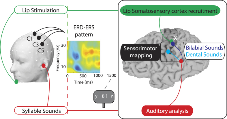
Figure 1. Experimental Design and Neurophysiological hypothesis.
On the left side a schematic of the experimental design: Lip Stimulation to the lower lip (which could be either present or absent) is shown in green and the Syllable Sound presentation (which could either be bilabial or dental syllables) is depicted in red. Electrodes within the left ROI (C1, C3, C5) are represented on the head (drawn by using EEGLAB functions72, version 13.4.4b, site URL http://sccn.ucsd.edu/eeglab/). In the center of the figure a Time-Frequency map of the ERSP, displaying the ERD and ERS pattern. Electrical stimulation to the lips and vowel’s onset are temporally aligned (red and green dashed lines at time = 0 ms). After 1500 ms a question regarding the heard sound appeared on the screen, requiring participants to respond using horizontal eye movements. On the right a schematic of the Neurophysiological Hypothesis: if speech is represented in a sensorimotor code (black box), each sound (after auditory analysis, in red) will be mapped onto distinct portions of the somatosensory cortex (post-central gyrus) depending of the specific place of articulation of the syllable (cyan or dark blue). Peripheral stimulation of the lips (in green) will match with one of the two somatosensory representations (bilabial sounds, dark blue). The white asterisk depicts the interaction between the processing of lip-related sounds with the lip-stimulation, due to the recruitment of overlapping neural resources (brain surface reconstructed from the MRI of one author using Freesurfer image analysis suite75, version 5.3.0, site URL http://freesurfer.net).

