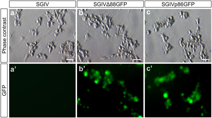
Figure 4. Pathogenicity of SGIV and its recombinant versions.
HXI cells were infected with SGIV, SGIV∆88GFP and SGIVp86GFP at MOI of 1. CPE and fluorescent signal were observed with microscopy. (a,a’) HX1 cells at 72 hpi with SGIV. (b,b’) HX1 cells at 72 hpi with SGIV∆88GFP. (c,c’) HX1 cells at 72 hpi with SGIVp86GFP. CPE is seen in bright field phase contrast micrographs (a–c) and GFP expression is shown in fluorescent micrographs (a’–c’). Scale Bars, 10 μm.

