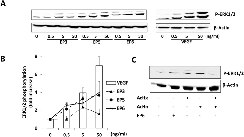
Figure 4. ERK1/2 pathway activation analysis.
(A) PAEC-VEGFR2 were incubated for 15 min at 37 °C in starvation medium with different concentration (0.5, 5 and 50 ng/mL) of EP3, EP5, EP6 peptides and VEGF. (B) Data in the graph report ERK1/2 phosphorylation increase respect to cells in serum deprivation condition (n = 3). (C) Comparison of the effect of EP6 and control peptides, tested alone and in combination, on ERK1/2 phosphorylation. PAEC-VEGFR2 were exposed to EP6 (50 ng/mL), AcHx (22 ng/mL), AcHn (26 ng/mL) and their combination for 15 min. Cytosolic extracts from treated cells were analyzed by Western blot using anti-phospho ERK1/2. Anti-β-actin antibody was used as loading control (western blot quantification expressed as fold increase respect to Ctr: EP6 3.8 ± 0.4, AcHx 3.1 ± 0.9, AcHn 2.9 ± 0.9, combination 1.9 ± 0.75).

