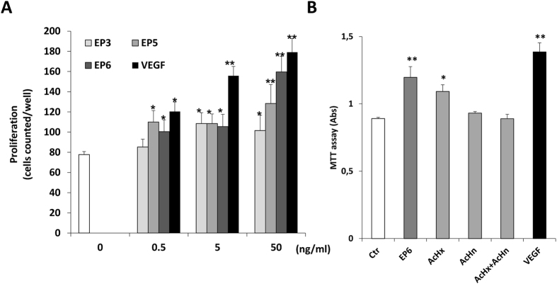
Figure 5. Effect of EP peptides on EC proliferation and survival.
(A) Subconfluent HUVEC cultures were incubated in starvation medium with the indicated concentrations of EP3, EP5 and EP6 for 72 h and then counted. VEGF was used as a positive control. P < 0.05 and **P < 0.01 vs basal control. (B) PAEC-VEGFR2 were stimulated with EP6 (50 ng/mL), AcHx (22 ng/mL), AcHn (26 ng/mL) and their combination, or VEGF (50 ng/mL) for 48 h. Cell survival was measured by MTT assay and data are reported as absorbance units (n = 3).

