
Figure 6. Effect of EP peptides on EC migration.
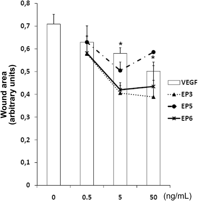
HUVEC monolayers were scratched with a pipette tip to mimic a wound. Adherent cells were then treated with increasing concentrations of EP3, EP5 and EP6 (0.5, 5 and 50 ng/mL), in the presence of the cell proliferation inhibitor ARA-C (2.5 μg/mL). VEGF was used as control. Wound area was measured after 18 h of incubation by image analysis. *p < 0.05 vs basal control. Data are expressed as arbitrary units of wound area taking as reference the area at time 0.