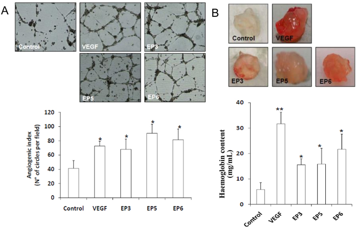
Figure 7. In vitro and in vivo angiogenic properties of EP peptides.
(A) HUVECs were plated onto a layer of basement membrane matrix (Matrigel) and incubated at 37 °C for 18 h in the presence of EP3, EP5, EP6 and VEGF (50 ng/mL). After treatment, photomicrographs of tubular structures (Top) were quantified as angiogenic index, calculated as the number of complete circles counted/field by microscope image analysis (Bottom). *p < 0.05 vs basal control. (B) In vivo Matrigel angiogenesis assay was performed by injecting Matrigel solution containing 500 ng of the selected EP peptides into the dorsal midline region of C57 black mice. Matrigel plugs loaded with VEGF (500 ng) or buffer were respectively used as positive and negative controls. After 10 days Matrigel implants were recovered (Top) and analyzed for their haemoglobin content (bottom). *p < 0.05 and **p < 0.01 vs Matrigel alone.

