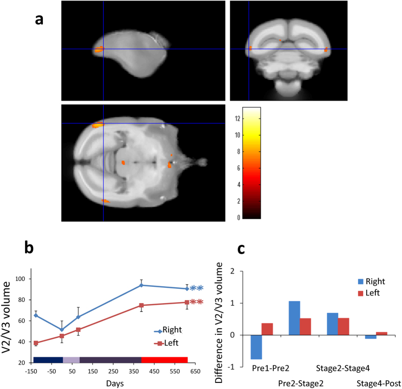
Figure 3. Gray matter increases in bilateral lateral extrastriate cortex (V2/V3).
(a) Bilateral gray matter increases in visual cortical area V3 and in left V2 as a function of experimental phases. (b) Time course of averaged gray matter change in peak voxels in ROI in bilateral V3, with SE (n = 4, **p < 0.01). Gray matter volume increased dramatically across the different training phases. Color bar on the bottom is the same as in Fig. 1. (c) Difference in bilateral V3 gray matter volume in both hemispheres between the adjacent periods, calculated per week. See also Fig. S2.

