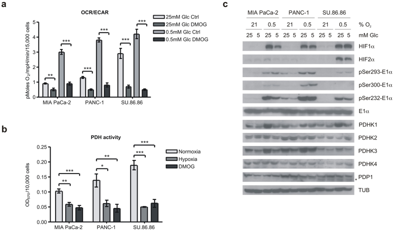
Figure 1. Hypoxia inhibits mitochondrial OCR and PDH activity and induces PDHK1 protein and activity.
(a) Ratio of oxygen consumption rate (OCR) to extracellular acidification rate (ECAR) measured by Seahorse XF in MIA PaCa-2, PANC-1, and SU.86.86 cell lines in high (25 mM) or low (0.5 mM) glucose incubated overnight with or without 1 mM DMOG. (mean ± SEM, two-tailed Student’s t-test, **p < 0.01, ***p < 0.001) (b) Cell-based PDH activity assay in cells incubated 16 h in normoxia, hypoxia (0.5% O2) or 1 mM DMOG. (mean ± SD, one-way ANOVA, *p < 0.05, **p < 0.01, ***p < 0.001) (c) Western blots of HIFα isoforms, pyruvate dehydrogenase kinase isoforms (PDHKs), phosphatase (PDP1 – lower band *), target phosphorylated serine residues on E1α and total E1α after overnight incubation in normoxia or hypoxia (0.5% O2) at 25 or 5 mM glucose as indicated.

