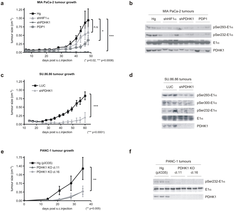
Figure 5. Decreased hypoxic response of PDH activity can slow the growth of xenografted tumours.
(a) Tumour growth curves of MIA PaCa-2 genetically modified cells (HIF1α and PDHK1 knockdown, PDP1 overexpression) described in Fig. 4 grown in nude mice (n = 8 per group in replicate experiments). Statistically significant differences exist between control (Hg) and shPDHK1 and PDP1 tumours as indicated. (mean ± SEM, two-way ANOVA) (b) Western blots from three random tumours of each group in (a) showing decreased PDHK1 expression and activity in vivo. (c) Tumour volumes of SU.86.86 control (LUC) and PDHK1 knockdown cells grown in nude mice (n = 8 per group). The growth rate differences were statistically significant. (mean ± SEM, two-way ANOVA) (d) Western blots from three random tumours in (c) showing decreased PDHK1 expression and activity in vivo. (e) Tumour volumes of PANC-1 control (Hg (pX335)) and 2 clones of PDHK1 null cells (PDHK1 KO) described in Fig. 2 grown in nude mice (n = 4 per group). The growth rate differences were statistically significant. (mean ± SEM, two-way ANOVA) (f) Western blots from three random tumours in (e) showing no PDHK1 expression and activity in vivo.

