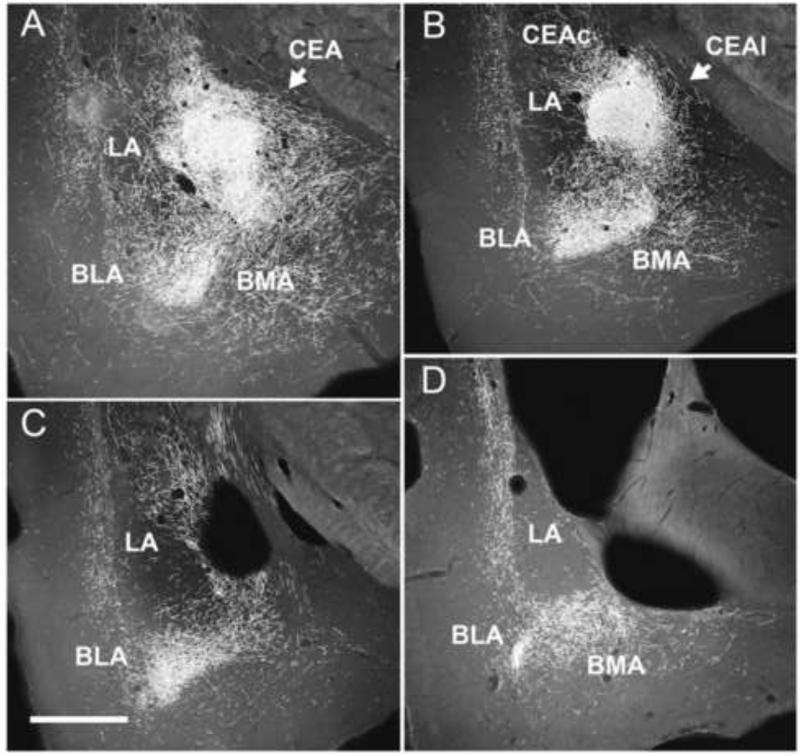
Figure 9.
A-D: Series of low magnification darkfield photomicrographs of transverse sections rostrocaudally (A-D) through the forebrain depicting patterns of labeling within the amygdala produced by a PHA-L injection in the posterior paraventricular nucleus of the midline thalamus. A,B: Note dense labeling in the central (CEA), basomedial (BMA) and basolateral (BLA) nuclei of amygdala, and marked but less pronounced labeling in parts of the medial, lateral (LA) and anterior cortical nuclei of the amygdala. C,D: Note prominent labeling caudally within the amygdala mainly confined to BMA and BLA. See list for abbreviations. Scale bar = 750 μm. Modified from Vertes and Hoover (2008).

